Deflector based dielectrophoretic separation of biomolecules using BioMEMS
The Project was successfully completed by Ms. Nivishna Venkatraj II year M.Tech and Dr. M.Ananthasubramanian (PI) Assistant Professor, Biotechnology from PSG College of Technology, Peelamedu, Coimbatore.

The project aims to Design the deflector based dielectrophoretic circular separator and to Test the model.
Dielectrophoresis (or DEP) is a phenomenon in which a force is exerted on a dielectric particle when it is subjected to a non-uniform electric field. This force does not require the particle to be charged. However, the strength of the force depends strongly on the medium and particles' electrical properties, on the particles shape and size, as well as on the frequency of the electric field. Consequently, fields of a particular frequency can manipulate particles with great selectivity. All particles exhibit dielectrophoretic activity in the presence of electric fields.
For a particle moving in a circular channel, different forces act on it, namely: the dielectrophoretic force and the fluid drag force. The resultant force in the radius direction acting on the particle at a distance r ,from the center is the sum of dielectric force and drag force. The particles with positive DEP feature will be driven away from the center. While the particles with negative DEP feature will be attracted towards the center. As a result, these particles with different DEP polarities will end up at the different outlets.
A Dielectrophoretic Bioparticle Seperator has multiple advantages which gives it an edge over the other methods of analysis. First advantage is that it is micro sized; the dielectrophoretic devices are table top easy to handle devices, it significantly reduces the time consumption. For a given pathogen analysis, carrying out conventionally using the culture methods would take at least seven days but using dielectrophoresis this can be accomplished in 15-20 minutes. Dielectrophoresis can be used for the separation of nanoscale particles like viruses, DNA, and proteins.
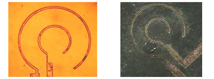
To conclude the dielectrophoretic bioparticle seperator was fabricated as per the proposed dimensions.The device was tested using a water droplet using a pulled pipette attached to a syringe. When a potential of 4-5V was applied to the device the water droplet disappeared due to the effect of electroysis or the heat generated caused the disappearance of the bubble.